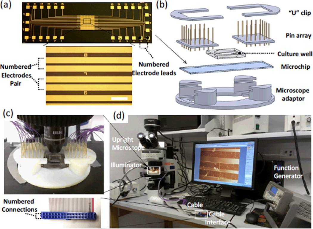
Fig. 2.
Experimental setup for selective electrical stimulation and cell contraction assessment: (a) Micropatterned interdigigated electrode array on a glass slide. Scale Bar=500µm. (b) Schematic showing the components of the selective stimulation device, including two U-shape clips, spring contact probes arrays, a cell culture well, an on-chip electrode stimulator and a microscope adaptor. (c) The assembled selective stimulation device was mounted on the stage of an upright microscope. (d) The experimental setup used for applying selective electrical stimulation and measuring cell contractile performance. The inset shows the cable interface with the numbered connections.

