
Figure 2.
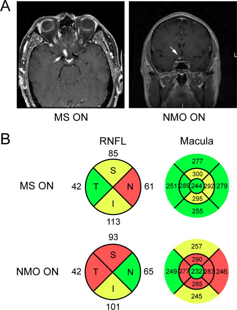
MRI and OCT imaging of NMO optic neuritis. A. Axial and coronal post-contrast T1-weighted images of acute NMO optic neuritis demonstrate acute inflammation of the prechiasmatic right optic nerve (arrows). B. Spectral domain OCT of the peripapillary RNFL and macula of eyes affected by MS- and NMO-associated ON. Note the increased retinal thinning in the macula affected by NMO ON despite a comparable amount of peripapillary RNFL loss (red: < 1st percentile; yellow: < 5th percentile; green: 5th–95th percentile). Thickness shown in microns.