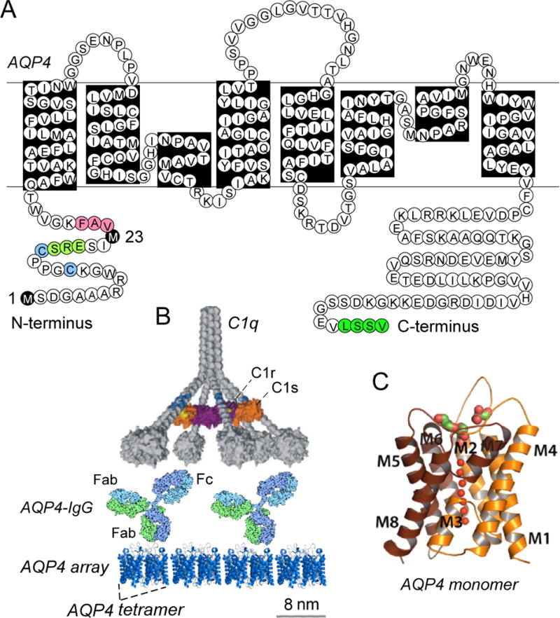
Figure 3.

AQP4, the target of AQP4-IgG autoantibodies. A. Amino acid sequence of human AQP4 showing Met-1 and Met-23 translation inhibition sites (black), residues involved in intermolecular N-terminus associations to form OAPs (pink); residues preventing OAP formation by M1-AQP4 (light green); cysteines involved in palmitoylation-regulated OAP assembly (blue); and C-terminus PDZ domain (dark green). B. CDC requires AQP4 assembly in OAPs. Multivalent binding of C1q to Fc regions of clustered AQP4-IgG on AQP4 OAPs. AQP4 tetramers shown with a generic IgG antibody and C1 on the same size scale. C. Crystal structure of human AQP4 (PDB, 3GD8).
