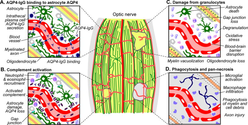
Figure 4.

Mechanisms of optic nerve damage in NMO. Lesions of different ages are depicted in a longitudinal section through myelinated retrolaminar optic nerve. A. Pre-lesion, showing AQP4-IgG accessing and binding to AQP4 on perivascular astrocytes. B. Acute early lesion, showing AQP4-IgG deposition and complement activation (purple discs, membrane attack complexes) and astrocyte damage. Cytokine release and recruitment of granulocytes into perivascular space is also shown. C. Subacute lesion, showing neutrophil and eosinophil degranulation that exacerbate astrocytotoxicity. Secondary oligodendrocyte loss results from disruption of glial tight junction networks and oxidative stress. Myelination is relatively preserved, albeit with prominent vacuolization. D. Older lesion, showing fragmentation and loss of myelin and appearance of activated macrophages and resident microglia. Axonal injury may arise from both failed remeylination and inflammatory damage.
