Figure 5.

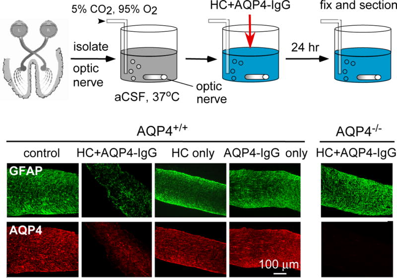
Ex vivo optic nerve culture model of NMO. Top, schematic showing mouse optic nerves cultured for 24 h in CO2/O2-bubbled artificial cerebrospinal fluid, with human complement (HC) and/or AQP4-IgG. Bottom, immunofluorescence for GFAP (green) and AQP4 (red) and myelin basic protein (MBP) (red) in wildtype (AQP4+/+) and AQP4 knockout (AQP4−/−) mice. ‘Control’ indicates no added AQP4-IgG or HC.
