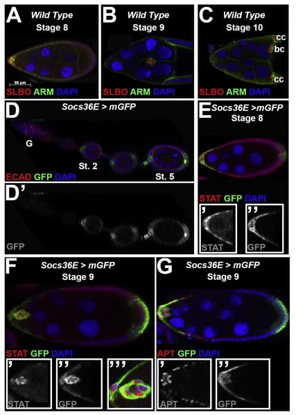
Fig. 1.
Socs36E expression in egg chambers overlaps with STAT activity during border cell specification and migration. For all images, anterior is to the left and the stages of oogenesis are indicated. (A–C) Wild type (Canton-S) egg chambers at mid-oogenesis are stained with antibodies directed against the border cell marker SLBO (red) and beta-cateninprotein (Armadillo, ARM, Green), which is also enriched in the border cells. DAPI (blue) labels DNA. (A) At stage 8, SLBO expression is graded in a subset of anterior-most follicle cells. (B) At stage 9, the border cells migrate between the nurse cells, and maintain SLBO expression. (C) The border cells (bc) reach the oocyte at stage 10, and SLBO is also expressed in centripetal cells (cc). (D–G) Socs36E-Gal4/UAS-mCD8-GFP egg chambersreveal the expression pattern of the Socs36Elocus. (D) GFP is not detected in the germarium (G) but is observed at stage 2 (St. 2) and later (Stage 5, St. 5) in a subset of anterior and posterior follicle cells. The inset (D) shows only GFP expression. (E–F) STAT protein expression, revealed by antibody staining (red), and the Socs36E reporter are both detected in the anterior follicle cells and border cell cluster atstages 8 (E) and 9 (F). The insets show the STAT (’) or GFP (”)staining, alone, or higher magnification at stage 9(’’’). (G) An antibody directed against APT (red) indicatesthis protein is detected in the same cells as GFP at stage 9. The insets show the APT (’) and GFP (”) stainings separately.

