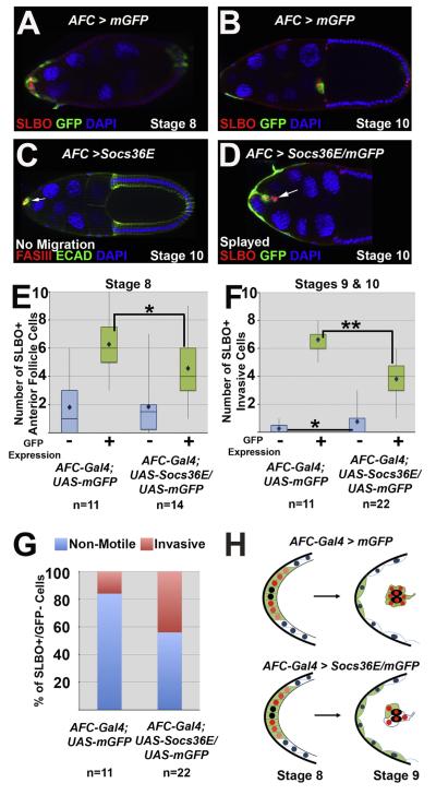
Fig. 2.
Ectopic Socs36E causes border cell identity, cohesion, and migration defects. For all egg chambers the stage of oogenesis and the mutant phenotype, if one is present, are indicated. “AFC”stands for Anterior Follicle Cells, where AFC-Gal4 indicates the lineP[GawB]c306. (A–B) AFC-Gal4; UAS-mCD8-GFP egg chambers at stage 8 (A) and stage 10 (B) indicate the expression pattern of the AFCdriver (by GFP antibody staining) in anterior follicle cells and the border cells, marked by SLBO antibody. (C) A stage 10 AFC-Gal4; UAS-Socs36E egg chamber, which over-expresses Socs36E in AFCs, stained with antibodies directed against FasIII (red) and E-Cadherin (green). A cluster of cells forms around the polar cells (indicated by FasIII) at the anterior end (arrow), but no cells migrate. (D) A stage 10 egg chamber co-expressing Socs36E andGFP in the presumptive border cell population(AFC-Gal4; UAS-Socs36E/UAS-mCD8-GFP). SLBO antibody (red) marks a small border cell cluster, which displays a splayed phenotype and fails to detach from the anterior end. The arrow indicates a pair of SLBO-positive/GFP-negative invasive cells. (E–F) Box and whiskers plots quantifyingSLBO-positive cells at stage 8 (E) and stages 9 and 10 (F) in egg chambers expressing GFP alone or with Socs36E under control of the AFC-Gal4 driver. For each genotype, the SLBO-positive cells are categorized as expressing GFP (green bars and +) or not (blue bars and −). The bars represent the range in the second and third quartiles, and mean cell counts are indicated by diamonds;(*) = p<0.05; (**) = p<0.0001. (G) Percent of the SLBO-positive, GFP-negative anterior follicle cells at stage 8 that become either invasive (pink) or remain as non-motile stretch cells (blue). (H) Schematics illustrating altered border cell recruitmentin wild type (top) or when Socs36E is over-expressed (bottom), as quantified in E and F. Polar cells are indicated by black, red nuclei indicate SLBO(+) cells, while blue nuclei are SLBO(−) cells. Green cells indicateGFP (+) cells, which express AFC-Gal4, while white cells do not.

