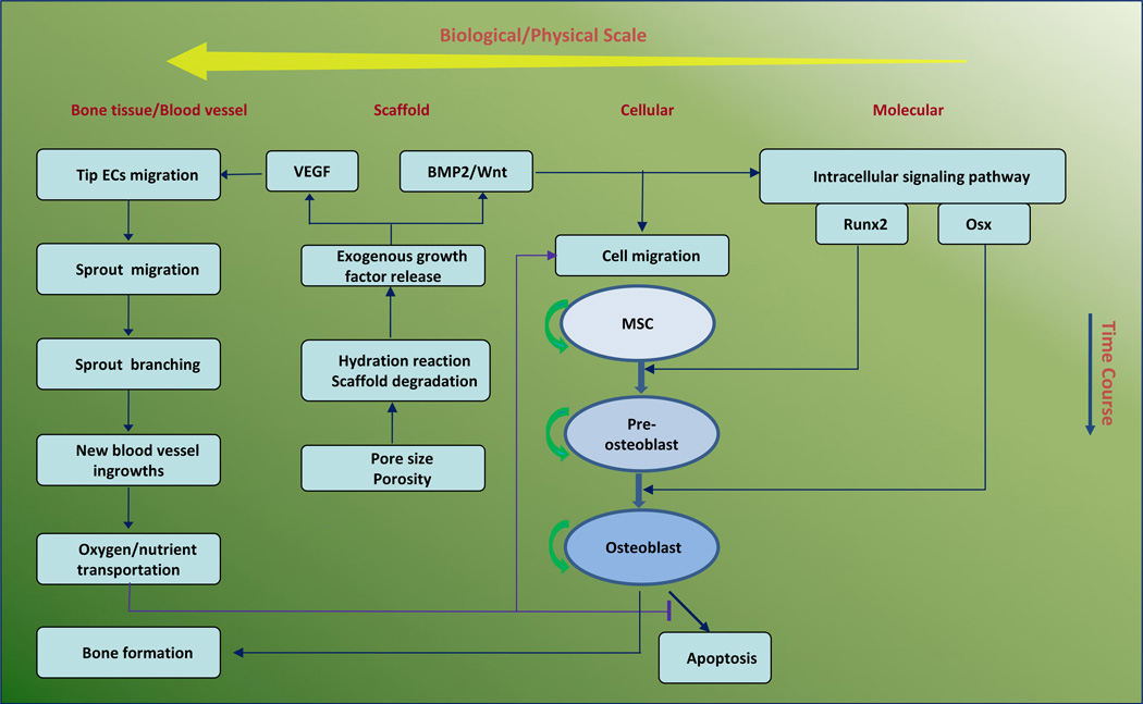
Fig. 1.
Schematic representation of computational framework of 3D vascularized bone regeneration within a porous biodegradable CaP scaffold. The model encapsulates four biological/physical scales from micro level to macro level: molecular, cellular, scaffold, and bone tissue scales. At the scaffold scale, growth factors (BMP2, Wnt and VEGF) were released from the CaP scaffold via calcium phosphate degradation due to hydration reaction. At molecular scale, the released BMP2 and Wnt stimulated the intracellular signaling pathway of MSC and pre-osteoblasts to activate the transcription factors Runx2 and Osx. At the cellular scale, each osteoblastic cell underwent migration, proliferation, differentiation, and apoptosis. MSC and pre-osteoblasts migrated along the gradient of the concentration of growth factors and oxygen, and their differentiation was regulated by Runx2 and Osx. At the bone tissue scale, new capillary sprouts, migrating from host tissue, were induced and sustained by released VEGF to grow into the pores of the scaffold. The remodelled vasculature could transport oxygen to maintain osteoblast metabolism and survival within scaffold pores.

