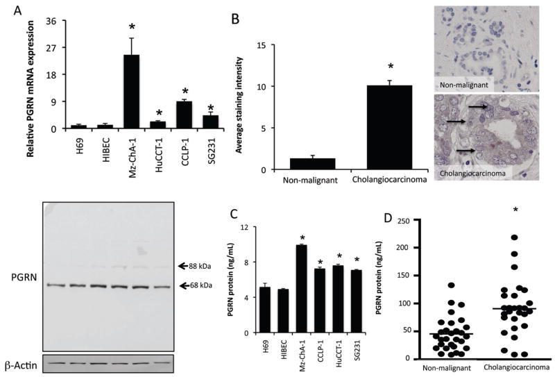
Figure 2.
PGRN expression and secretion is increased in cholangiocarcinoma. PGRN levels were assessed in four cholangiocarcinoma cell lines as well as non-malignant cholangiocyte cell lines H69 and HIBEC by real time PCR and immunoblotting (A). For real time PCR, data are expressed as average ± SEM (n=4). (*P<0.05 compared to PGRN in H69 cells). Representative PGRN immunoblots are shown (lower panel); β-Actin is shown as a loading control. PGRN levels were also assessed in biopsy samples from 48 cholangiocarcinoma patients and non-malignant controls by immunohistochemistry. Representative photomicrographs of PGRN immunoreactivity are shown (B; magnification ×40). Staining intensity was assessed as described in the methods and expressed as an average ± SEM of all cholangiocarcinoma patients compared to control samples (B; *P<0.05 compared with PGRN immunoreactivity in control biopsy samples). PGRN levels in the supernatant of cell suspensions of cholangiocarcinoma cell lines and the non-malignant cholangiocyte cell lines H69 and HIBEC were determined by EIA after 6 hr (C). Data are expressed as average PGRN concentration (ng/mL) ± SEM (n=3; *P<0.05 compared with PGRN levels secreted from H69 cells). PGRN levels in bile samples from cholangiocarcinoma and intrahepatic cholelithiasis patients were assayed by EIA (D). Data are expressed as average PGRN concentration (ng/mL) ± SEM. (reprinted with permission from Frampton, et al. Gut, 2012;61:268–77) (43)

