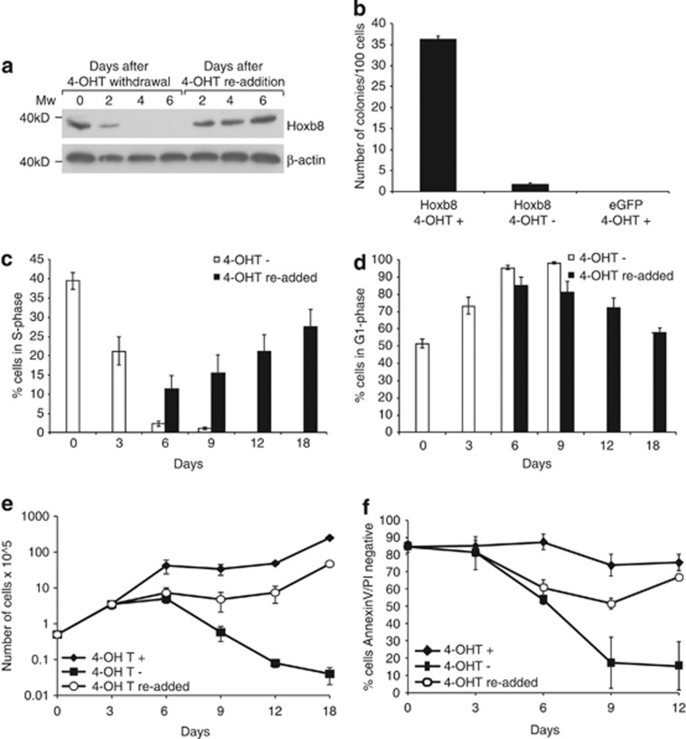
Figure 1.
Hoxb8 FDM cells stop proliferating and undergo apoptosis in the absence of Hoxb8 expression. (a) Lysates from Hoxb8 FDM cells were prepared at the indicated times after 4-OHT withdrawal and following 4-OHT re-addition after 4 days of withdrawal. Membranes were probed with antibodies to detect Hoxb8 and beta-actin as a loading control. (b) Hoxb8 expression is required for colony formation. Viable c-kit+ve/lin−ve cells were infected with Hoxb8 or eGFP encoding lentivirus, in the presence (+) or absence (−) of 4-OHT. Infected cells were single-cell sorted into 96-well plates and the number of colonies counted 14 days later. Results represent means±S.E.M. of four independent infections of four independent pools of c-kit+ve/lin−ve cells. (c) Cells in S-phase diminish after Hoxb8 downregulation. Hoxb8 FDM cells were cultured in IL-3 without 4-OHT (−) or following re-addition of 4-OHT after a 3-day period of withdrawal (re-added). At the indicated time points, cell-cycle analysis was performed using hypotonic PI buffer staining and flow cytometric analysis. The percentage of cells in S-phase was determined using the cell-cycle analysis software ModFit. Results indicate means±S.E.M. of seven independent clones in three independent experiments. (d) Cells arrest in G1 after Hoxb8 downregulation. Hoxb8 FDM cells were prepared as described in (c) and the percentage of cells in G1 phase was determined using Modfit. Results indicate means±S.E.M. of seven independent clones in three independent experiments. (e) Hoxb8 expression maintains proliferation in IL-3. Hoxb8 FDM cells were cultured in IL-3 with 4-OHT (+), without 4-OHT (−) or following re-addition of 4-OHT after a 3-day period of withdrawal (re-added). At the indicated time points, cell number was counted (see Materials and Methods). Results are means±S.E.M. of seven independent clones in three independent experiments. (f) Hoxb8 expression maintains viability in IL-3. Cell viability was determined by PI exclusion and FITC-conjugated AnnexinV staining from the same samples as described in (e). Results are means±S.E.M. of seven independent clones in three independent experiments

