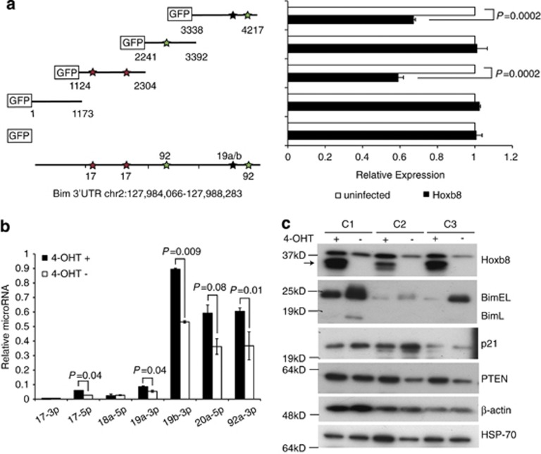
Figure 5.
Bim 3′UTR segments, which contain miR-17, miR-19a/b and miR-92 binding sites, are required for Hoxb8-mediated repression of Bim. (a) Murine Bim 3′UTR reporter activity in segments containing binding sites for miR-17, miR-19a/b and miR-92. 293T cells stably expressing the indicated GFP reporter plasmids or GFP alone were left uninfected or infected with Hoxb8 and GFP fluorescence analysed by flow cytometry. Fluorescence is relative to the uninfected cell population for each GFP reporter construct. Results are mean±S.E.M. of three independent experiments. P-values were derived using Student's t-test (two-tailed, equal variance). The star symbols indicate miRNA binding sites shown in the representation of the full Bim 3′UTR at the bottom of the panel. These sites were derived from the target prediction software, TargetScan, and are representative of a number of miRNAs with identical seed regions. These include miR17-5p/20ab/20b-5p/92/106ab/427/518-3p/519d for the miR-17 site and miR25/32/363/363-3p/367 for the mir-92 site. (b) qRT-PCR analysis of expression levels of miR-17-3p, miR-17-5p, miR-18a-5p, miR-19a-3p, miR-19b-3p, miR-20a-5p and miR-92a-3p in wild-type Hoxb8 FDM cells in the presence (+) and 4-day absence (−) of 4-OHT. All miRNA levels are normalised to U6 and expressed relative to miR-16. Results are mean±S.E.M. of three independent clones analysed in triplicate in two independent experiments. P-values were derived using Student's t-test (two-tailed, equal variance). (c) Western blot of other predicted miR-17∼92 targets in Hoxb8 FDM cells. Lysates of Hoxb8 FDMs cultured in the presence (+) and 4-day absence (−) of 4-OHT were analysed by western blotting for antibodies against Hoxb8, Bim, p21 and PTEN. Beta-actin is the loading control for all antibodies excluding PTEN for which HSP-70 is shown. Three individual clones, C1, C2 and C3, are shown. Arrow indicates Hoxb8

