Figure 7.
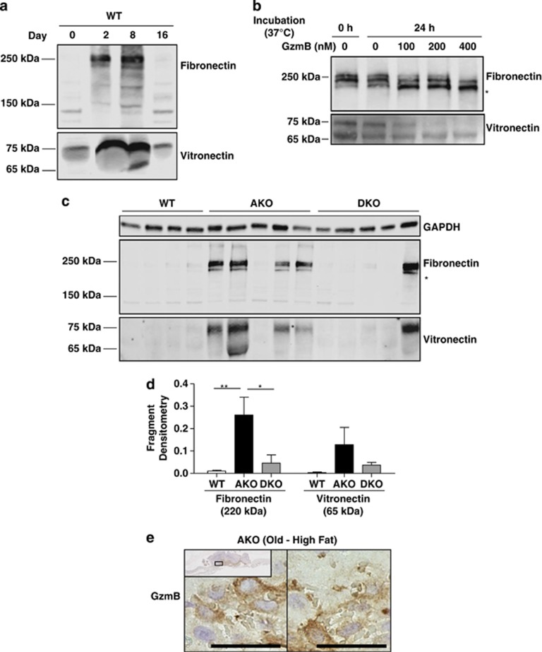
Fibronectin and vitronectin content during wound healing. (a) Wounded skin from WT mice was analyzed at days 0, 2, 8 and 16 for fibronectin and vitronectin by western blot. Before wounding (day 0), both fibronectin and vitronectin content is relatively low. Beginning from day 2 to day 8, granulation tissue forms, featuring an increase in fibronectin and vitronectin that returns to normal on day 16. (b) Mouse GzmB was added to the skin homogenate from a WT mouse harvested at day 8 post-wounding. As little as 100 nM of GzmB was sufficient to degrade the full-length fibronectin and vitronectin proteins and generate a fibronectin fragment at ∼220 kDa. (c) AKO mice exhibited increased fibronectin and vitronectin content including the ∼220 kDa fibronectin fragment, which appeared to be reduced but not eliminated by knocking out GzmB. (d) The 220-kDa fibronectin fragment and the 65-kDa vitronectin fragment were quantified by densitometry. (e) GzmB staining was evident within the granulation tissue of HFD-fed AKO mice at day 16. Both images taken from the same wound (inset shows the low magnification image). *P<0.05 and **P<0.01, two-way ANOVA with Bonferonni post test. Scale bars=25 μm

