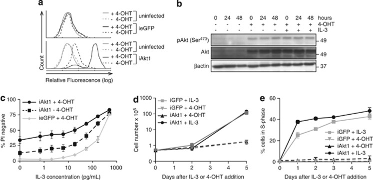
Figure 2.
Enforced expression of constitutively active Akt increases survival of FDM cells in low IL-3 concentrations. (a) Expression of inducible Akt1 as detected by intracellular FACS analysis using FITC-conjugated anti-HA antibody. Representative histograms of Akt expression compared with uninduced and ieGFP expressing cells are shown. (b) Lysates of iAkt1 expressing cells cultured in the presence or absence of 4-OHT and IL-3 were probed with antibodies to detect phosphorylated Akt (serine 473), total AKT and β-actin as a loading control. (c) The viability of multiple independent WT clones of FDM cells expressing inducible myr-HA-(ΔPH)Akt1 (iAkt1; n=4) cultured in the presence or absence of 4-hydroxytamoxifen (4-OHT) was compared with inducible GFP (ieGFP) expressing cells cultured in the indicated concentrations of IL-3 for 72 h. The results represent means and standard error of the mean of three independent experiments. (d and e) FDM cells expressing iAkt1 were starved of IL-3 overnight, and either IL-3 or 4-OHT added, the later to induce expression of activated Akt. Cell number was counted using haemocytometer (d) and the proportions of cells in S phase (e) were determined by using intracellular PI staining and flow cytometry over a 5-day period

