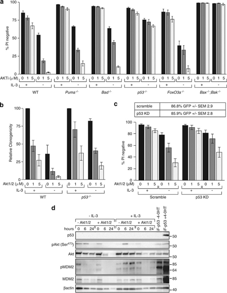
Figure 6.
Apoptosis induced by Akt inhibition requires p53 but not Puma. (a) FDM cells of the indicated genotypes were cultured in the presence or absence of IL-3 and the AKT inhibitor (AKT1/2) at the indicated doses. Cell viability was measured by PI exclusion by flow cytometry. The numbers of independent clones (n) tested were as follows: WT n=5, Puma−/− n= 3, Bad−/− n=2, p53−/− n=3, FoxO3a−/− n=3, Bax−/−;Bak−/− n=2. The results represent means±S.E.M. of all clones tested in at least two independent experiments. Not all clones were tested in all experiments. (b) Multiple WT clones (n=4) or p53−/− clones (n=3) of FDM cells were cultured as in (a) before being washed and replated into medium containing soft agar and IL-3 (0.5 ng/ml). The numbers of colonies were counted after 10 days and expressed relative to the numbers of colonies in cultures not exposed to AKT1/2. Data represent means and standard errors of three independent experiments. (c) WT clones were transduced with retroviral constructs encoding a p53 shRNA or a scrambled control. Infected cells expressed GFP and the percentage of infected cells (±S.E.M.) is shown in the table. Viability was determined by PI exclusion in the presence or absence of IL-3 and the indicated doses of Akt inhibitor (AKT1/2). Data are means±S.E.M. of three independent clones tested in two independent experiments. (d) Western blot of WT FDM cells cultured in the presence or absence of IL-3 and the absence or presence of 5 μM AKT1/2 for 0, 6 and 24 h were probed with antibodies to detect p53, pAkt (Ser473), total Akt, MDM2, pMDM2 and β-actin as a loading control. WT FDM clone infected with lentiviral construct encoding FLAG-tagged p53 under the control of a 4-OHT inducible promoter, in the presence and absence of 4-OHT, is shown as a control for p53 expression

