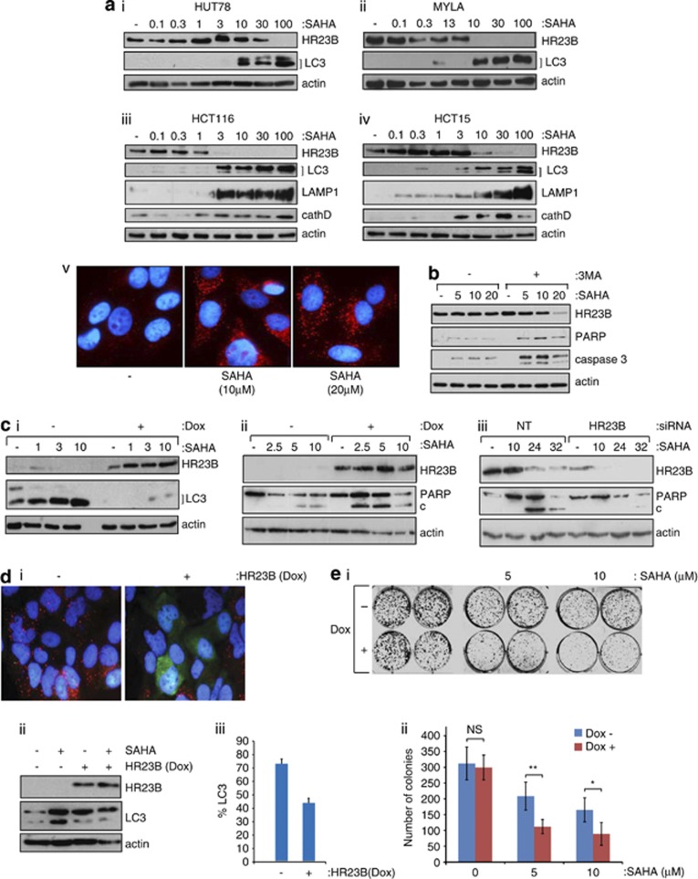
Figure 1.
Autophagy and HR23B in cells treated with HDAC inhibitors. (a)HUT78(i) and MYLA(ii) (CTCL), and HCT116(iii) and HCT15(iv) (CRC) cells were each treated with SAHA at the indicated (μM) concentrations, extracts prepared at 18 h and immunoblotted with anti-HR23B, anti-LC3, anti-LAMP1, anti-cathepsin D or anti-actin antibody as indicated; note that the appearance of the lower form (LC3-II) and increased overall LC3 level is indicative of autophagy. Visualisation of autophagosomes in U2OS cells by immunostaining with anti-LC3 (in red) in SAHA (10 and 20 μM) treated cells is also shown (v). Counter-stain with DAPI was superimposed for each image. (b) U2OS cells were treated with SAHA at the indicated (μM) concentration together with 3-methyladenine (3-MA; 10 mM) for 18 h, when cell extracts were immunoblotted with the indicated antibodies. (c) (i) U2OS cells stably expressing inducible HR23B were either untreated or treated with SAHA as indicated (in μM for 18 h) under non-induced (−) or induced (+) doxycycline treatment conditions (1 μg/ml; induction for 72 h treatment). Cells were harvested and immunoblotted with anti-Flag (for ectopic HR23B), anti-LC3 or actin as indicated. (ii) U2OS stable cells were treated as described in (i) with SAHA (at the indicated concentrations in μM) for 24 h, and the level of apoptosis analysed by immunoblotting for PARP (cleaved (c) and uncleaved) under non-induced (−) or induced (+) doxycycline treatment conditions as indicated. The level of ectopic HR23B and actin is shown. (iii) U2OS cells were treated with HR23B or control (NT) siRNA (50 nM for 72 h) in the absence (−) or presence of SAHA (10 μM) for the indicated times (in h) and the level of PARP (cleaved (c) and uncleaved) measured. The level of HR23B and actin is shown. (d) (i) Autophagosomes in U2OS cells visualised with anti-LC3 (red) stably expressing inducible HR23B (visualised with anti-Flag; green) after treatment with SAHA (10 μM for 18 hr) in the absence (−) or presence (+) of doxycycline. (−) shows the LC3 staining autophagosomes (red) under SAHA treatment, and (+) shows superimposed LC3 (red) and HR23B (green) images. (ii) The levels of HR23B and LC3 were analysed by immunoblotting with anti-Flag (ii), and quantitation of cells with autophagosomes after treatment with SAHA is presented in (iii), in the absence (−) or presence (+) of doxycycline, which was performed in triplicate (error bars indicate S.E.M.). (e) (i) U2OS cells stably expressing HR23B were grown in duplicate in the absence (−) or presence (+) of doxycycline together with SAHA (indicated concentration in μM) and, after 9 days, the number of viable cell colonies assessed by crystal violet staining. The untreated control cells are shown for comparison. (ii) Graphical representation of the data in (i), where values indicate mean ±S.E.M; n=3; *P<0.05 and **P<0.01 Student's t-test

