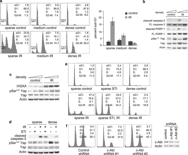
Figure 1.
DNA damage-induced apoptosis is inhibited at high cell density. (a) MCF10A cells were plated at different cell densities. Cells were irradiated at 20 Gy, and were harvested for fluorescence-activated cell sorting (FACS) analysis 72 h post-irradiation (IR). Left panels, profile plots of representative FACS analysis. Percentages of cells in the different fractions (sub-G1, G1, S and G2-M) are indicated. Right panel, graph showing quantification of three independent experiments. (b) Cells were treated as in (a), and analyzed by immunoblot with the indicated antibodies. (c) Cells were plated and irradiated as in (a), but were harvested 2.5 h post-IR for immunoblot analysis. (d) NIH3T3 cells were plated sparse or dense and were treated with 10 μM STI571 or dimethyl sulfoxide (solvent control) as indicated. Cells were irradiated 2.5 h later at 40 Gy, and were harvested 96 h post-IR, and analyzed by immunoblotting. (e) NIH3T3 cells were treated as in (d), and were analyzed by FACS. Percentages of cells in the different fractions are indicated. (f) MCF10A stably expressing small hairpin RNA (shRNA) to c-Abl or non-targeting control were plated sparse, and were irradiated at 25 Gy. Left: cells were harvested for FACS analysis 96 h post-IR. Percentages of cells in the different fractions are indicated. Right: the expression of c-Abl in the c-Abl-shRNA-expressing cells was analyzed by immunoblotting

