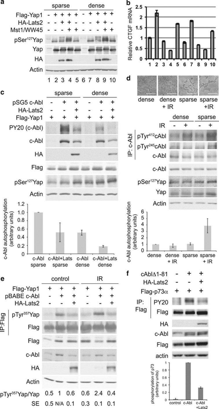
Figure 3.
High-cell density inhibits c-Abl autophosphorylation. (a–b) Activation of Lats by overexpression and high-cell density. HEK293 cells were transfected with the indicated and green fluorescent protein (GFP)-expressing plasmids. Cells were replated 28 h post-transfection to produce sparse and dense plates. Sixteen hours later, cells were harvested and immunoblotted with the indicated antibodies (a). (b) CTGF mRNA levels were analyzed by qPCR and normalized by GFP mRNA. (c) High cell density and Lats2 overexpression inhibit c-Abl autophosphorylation. HEK293 cells were transfected with Flag-Yap1 (all lanes), and indicated plasmids. Transfected cells were replated sparse and dense, harvested 11 h later, and analyzed by immunoblotting. PY20 detects phosphotyrosine. Lower panel: Quantification of c-Abl autophosphorylation, N=2. (d) c-Abl DNA damage-induced activation is inhibited at high cell density. HEK293 cells were plated at high and low cell density. Cells were irradiated at 20 Gy, and were photographed (upper panel, scale bar=0.1 mm) and then harvested 24 h post-IR. Middle panel: c-Abl was immunoprecipitated and samples were analyzed by immunoblotting with the indicated antibodies. Lower panel: Quantification of c-Abl autophosphorylation, N=6. (e) Lats2 inhibits IR-induced c-Abl phosphorylation of Yap1. HEK293 cells were transfected with the plasmids indicated, and 24 h post-transfection, cells were irradiated at 20 Gy. Cells were harvested 4 h post-IR, and Flag-Yap1 was immunoprecipitated. Samples were analyzed by immunoblotting with the indicated antibodies. Quantification of pTyr357 Yap, N=3. (f) Lats2 inhibits c-Abl phosphorylation of p73. Proteins were immunoprecipitated from transfected HEK293 cells with anti-Flag. Upper panel: Samples were analyzed by immunoblotting with the indicated antibodies. Lower panel: Quantification of p73 phosphorylation, N=3

