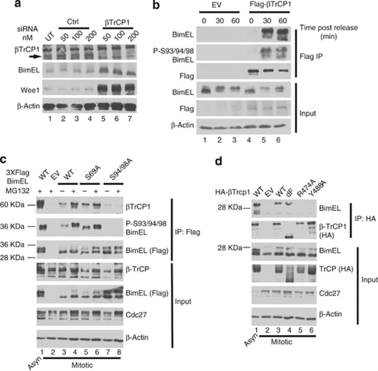
Figure 4.
Phosphorylation on Serine 93/94/98 of BimEL creates a binding site for βTrCP1. (a) Knockdown of βTrCP1 expression in HeLa cells using siRNA at the indicated concentrations. A non-silencing siRNA (Ctl) was used as control. Immunoblot analysis was performed against endogenous BimEL and Wee1. β-Actin was monitored as loading control. (b) HeLa cells were transfected with Flag-tagged βTrCP1 or empty vector control (EV). Cells were synchronized in mitosis as in Figure 1a except that cells were released in the presence of MG132. Cell lysates prepared at the indicated times post release. Immunoprecipitation (IP) was performed using anti-Flag antibody and immunoblot analysis used to detect endogenous BimEL, Flag (βTrCP1), and phosphorylated BimEL on serines 93/94/98 (P-S93/94/98). Cell extracts used as input for IP were analyzed by immunoblot to measure expression of total BimEL, Flag (βTrCP1), and β-Actin as loading control. (c) 293T cells were transfected with EV, wild-type (WT) BimEL or phosphorylation site mutants as indicated. Cells were synchronized in mitosis as in Figure 1a. Asynchrous (Asyn) cells treated with PMA and MG132 was used as a positive control (lane 1). Left: asynchronous (Asyn). IP was performed using anti-FLAG antibody and immunoblot analysis used to detect endogenous βTrCP1, Flag (BimEL), and phosphorylated BimEL on serines 93/94/98 (P-S93/94/98). Cell extracts used as input for IP were analyzed by immunoblot to measure expression of total βTrCP1 and Flag (BimEL). Cdc27 immunoblots were used to confirm mitotic state of cells and β-actin as loading control. (d) 293T cells were transfected with EV, wild-type (WT) βTrCP1 or the indicated βTrCP1 mutant. Cells were synchronized as in Figure 1a. Asynchronous cells transfected with HA bTrCP1 WT and treated with PMA and MG132 for 3 h was used as a positive control (lane 1). IP was performed using anti-HA antibody and immunoblot analysis used to detect IPed BimEL and HA (βTrCP1). Cell extracts used as input for IPs were analyzed by immunoblot to measure expression of total Bim and HA (βTrCP1). Cdc27 immunoblots were used to confirm mitotic state of cells and β-actin as loading control

