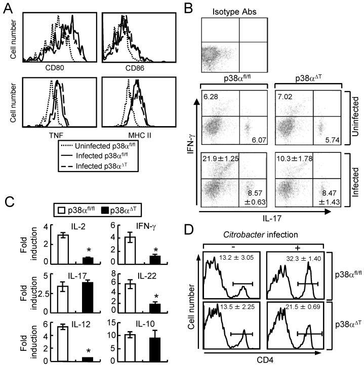
Figure 3. p38α promotes the activation and recruitment of T cells to the site of C. rodentium infection.
(A) Activation of DC is not affected in p38αΔT mice. DCs from MLN of C. rodentium-infected p38αfl/fl or p38αΔT mice were obtained after 1 week of infection, and stained with the indicated antibodies for FACS analysis. DCs from uninfected p38αfl/fl were used as the control. (B) Inflammatory cytokine expression in LP T cells. LP lymphocytes from C. rodentium-infected p38αfl/fl or p38αΔT mice were obtained after 1 week of infection, and stained with anti-CD3-PerCP and anti-CD4-APC Abs. Cells were further permeabilized and stained with anti-IL-17A-FITC and anti-IFN-γ-PE Abs. LP T cells from uninfected mice were used for the staining of isotype antibodies. The numbers indicate the percentage of the cells. (C) The mRNAs levels of inflammatory cytokines of enriched T lymphocytes from LP were measured by the quantitative PCR. Cells were obtained from uninfected or infected mice after 1 week of C. rodentium infection, and total RNA was prepared for quantitative PCR. Values are shown as the fold changes of each gene from the cells of infected mice compared to the uninfected control. Actin level was measured as an internal control. *, p<0.05, and error bars indicate s.d. (D) Recruitment of T cells to the LP. LP lymphocytes were obtained from uninfected mice or mice after 1 week of infection, and the proportion of CD4 cells was analyzed by FACS analysis. Numbers indicate the percentage of CD4-positive cells. Data are representatives of 2-3 experiments.

