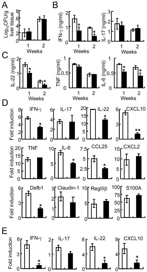
Figure 4. Function of intestinal epithelial cells (IECs) was affected by reduced activation and recruitment of T cells in C. rodentium-infected p38αΔT mice.
(A) Epithelial cell integrity is normal. Liver tissues were obtained from C. rodentium-infected p38αfl/fl (□) or p38αΔT (■) mice after 1 or 2 weeks of infection, and C. rodentium CFU was measured. (B &C) Ex vivo culture of IECs to measure the production of inflammatory cytokines. Colon tissues of p38αfl/fl (□) or p38αΔT (■) mice were obtained after 1 week of C. rodentium infection, and incubated for 24 hours. Levels of cytokines in culture supernatants were measured by ELISA. (D & E) Expression of inflammation genes in the colon tissues (D) or purified IECs (E) of C. rodentium-infected p38αfl/fl (□) or p38αΔT (■) mice. Total RNAs were obtained from colon tissues or IECs after 1 week of infection. RNA samples were also prepared from uninfected mice as a control. Quantitative PCR analysis was performed to measure the expression of target genes. Values are shown as the fold changes of each gene in infected samples compared with the uninfected control. Actin level was measured to normalize the values. *, p<0.05, and error bars indicate s.d. Results are representatives of 2-3 experiments.

