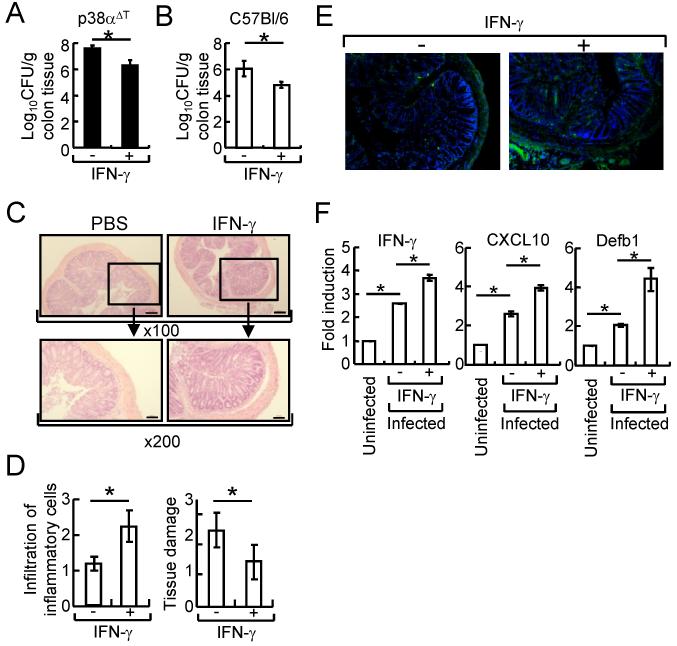
Figure 5. Administration of IFN-γ enhanced host defense activity of C. rodentium-infected mice.
(A) p38αΔT mice were orally inoculated with C. rodentium, and intraperitoneally injected with IFN-γ (10 μg/mouse) or PBS on days 0, 2, and 4 after infection. C. rodentium CFU in colon tissues was measured after 10 days. (B-F) The same as (A) except wildtype mice were used and colon tissues were obtained after 10 days. Bacterial counts in colon tissues were measured (B). (C & D) H & E staining (C) and histological scoring of the infiltration of inflammatory cells and tissue damage (D, n=6) was assessed. Boxed area is X200 of the original X100 magnification. Scale bar = 20 μm. (E) Recruitment of T cells was examined by immunostaining using anti-CD4 Ab (green). Nuclei were counterstained with DAPI (blue). Original magnification X100. (F) Expression of IFN-γ, CXCL10, and Defb1 in IECs. Actin level was used as an internal normalization control, and fold induction of each gene was compared to that of uninfected mice. *, p<0.05, and error bars indicate s.d. Representative results of 2-3 experiments are shown.

