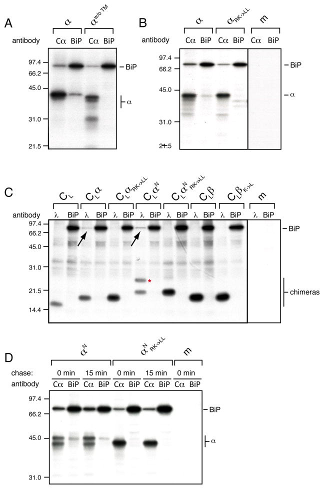
Figure 3. BiP binds the TM region of ER-lumenal α-chains.
(A) Metabolically labeled lysates from COS-1 cells transfected with the full-length α-chain construct (α) or one devoid of its TM region (αw/o ™) and BiP were split and immunoprecipitated with either an anti-Cα antibody or anti-BiP antiserum and analyzed by SDS-PAGE.
(B) COS cells co-transfected with BiP and either the wild type α-chain construct (α) or one in which the TM basic residues were mutated to leucine (αRK->LL) were analyzed as in (A).
(C) Interaction of radiolabeled BiP with CL-based reporter constructs (see Figure 1). Metabolically labeled lysates of cells co-transfected with BiP and one of the indicated constructs were divided and immunoprecipitated with either anti-mouse λ antiserum or anti-BiP antiserum. The construct glycosylated at its C-terminal reporter site is marked with a red asterisk. Co-immunoprecipitating BiP is indicated with a black arrow.
(D) Analysis of the interaction of αN and αNRK->LL with BiP. Cells co-transfected with the indicated constructs and BiP were subjected only to a 30 min pulse (0 min chase) or chased for an additional 15 min prior to cell lysis and immunoprecipitation with the indicated antibodies (m: mock transfection). See also Figure S3.

