Fig. 2.
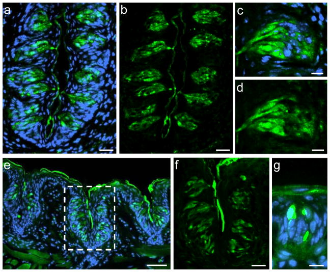
Visualization of GPR92 in murine taste papillae. Immunolabeling of cross sections through the murine circumvallate papillae (a–d), the foliate papillae (e–f) or the fungiform papillae (g).
(a, b) Numerous GPR92-positive cells are located within taste buds of the circumvallate papilla.
(c, d) Magnification of a circumvallate taste bud. GPR92-positive cells show the typical, spindle-shaped morphology of taste cells.
(e) GPR92-immunoreactive cells in taste buds of foliate papillae.
(f) Magnification of the dotted area in (e). GPR92-immunoreactive cells in foliate taste buds.
(g) GPR92-positive cell in a fungiform papilla.
Sections are counterstained with DAPI (blue). Scale bars: a, b, f = 20 μm; c, d, g = 10 μm; e = 50 μm

