Fig. 4.
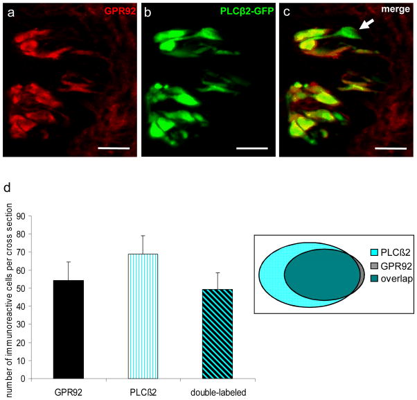
GPR92 is expressed in type II taste cells.
Immunohistochemistry employing an GPR92 antibody on cross sections through the circumvallate papilla of a PLCβ2 transgenic mouse.
(a) GPR92-immunoreactive cells in circumvallate taste buds.
(b) Numerous GFP-positive cells in the same circumvallate taste buds.
(c) The overlay of (a) and (b) clearly reveals that the vast majority of GPR92-immunoreactive cells is also GFP-positive. However, a subset of GFP-expressing cells shows no labeling for GPR92 (arrowhead).
(d) Quantitative analyses of co-expression patterns. GPR92-positive cells, PLCβ2-GFP-positive cells and double-labeled cells (GPR92-positive and PLCβ2-GFP-positive) in the circumvallate papilla were counted on cross sections. The analyses revealed that 91.4% of GPR92-immunoreactive cells are GFP-positive. In contrast, 72.0% of GFP-expressing cells show GPR92-immunoreactivity. Twenty sections from the circumvallate papilla of one mouse were analyzed. Data are expressed as mean numbers ± SD. Sections are counterstained with DAPI (blue). Scale bars: a–c = 20 μm

