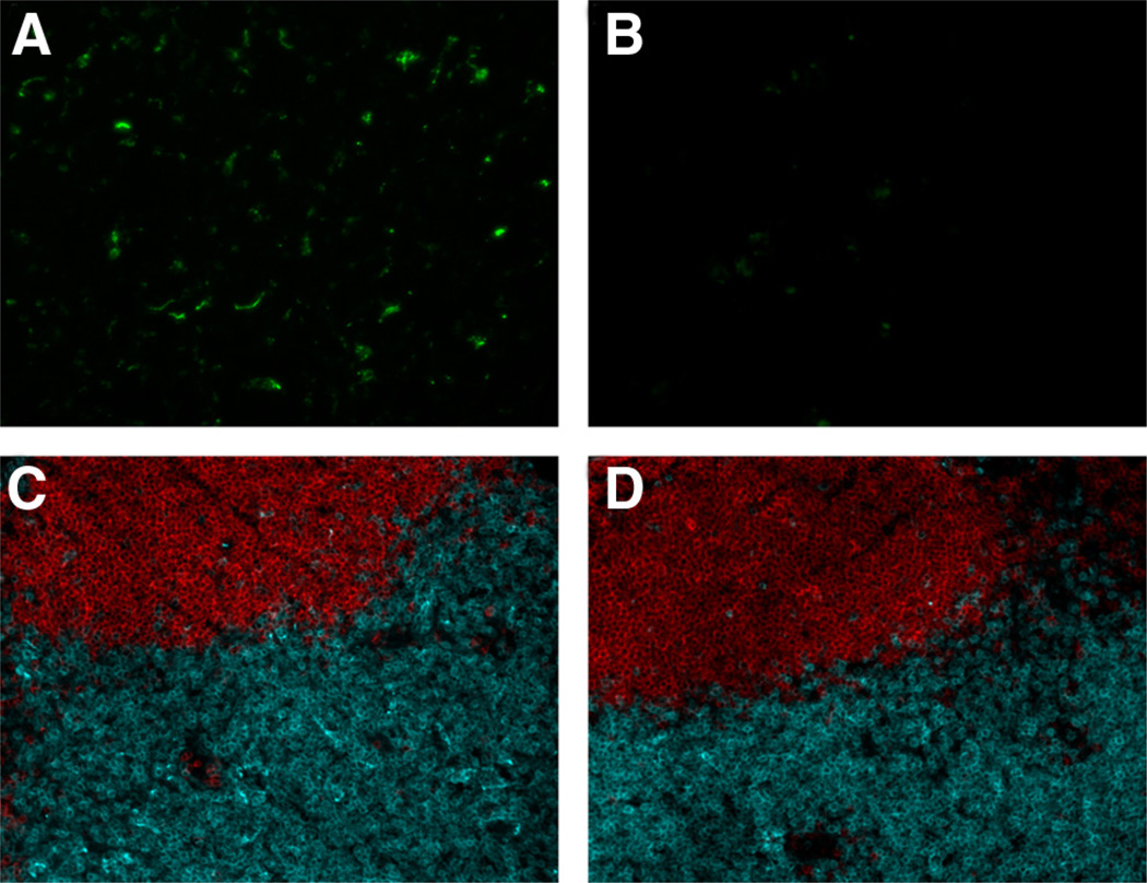
FIGURE 5.
Treatment of CD11c-DTR mice with DT causes depletion of GFP+ cells in lymph nodes but does not affect normal lymph node architecture 18 h after treatment. CD11c+ cells are revealed by GFP expression in CD11-DTR mice treated with PBS (A). The level of GFP detected was greatly reduced in mice treated with DT (B). Sections stained with CD19-specific (red) and CD3-specific (blue) Abs revealed no difference in normal lymph node architecture in T and B lymphocyte zones between PBS control mice (C) and DT-treated mice (D). The data in this experiment were obtained from four mice per group.

