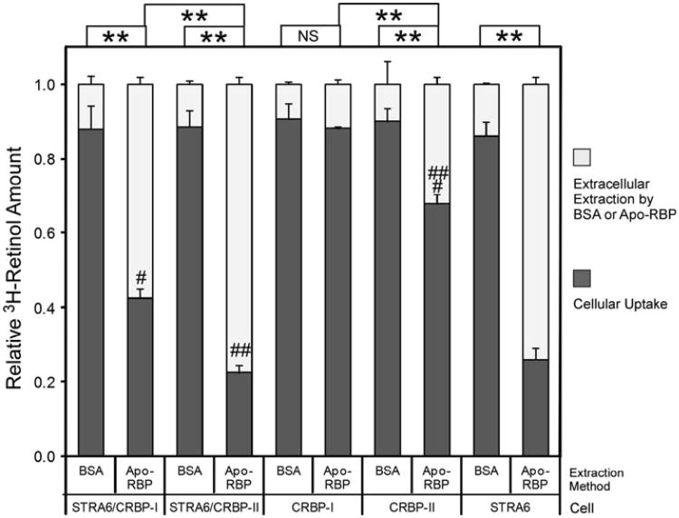
Fig. 3.
Comparison of CRBP-I and CRBP-II in STRA6-mediated retinol efflux. After cells were loaded with free 3H-retinol for 2 h, 1 μM of apo-RBP was added to cause 3H-retinol depletion for 2 h. BSA (1 μM) of was used as a control. 3H-retinol extracted during the 2-h incubation with apo-RBP or BSA is shown in the white column. 3H-retinol remaining in the cells after extraction is shown in the dark gray column. The total amount of 3H-retinol taken up by cells before the extraction is represented by combining the white column and dark gray column and is defined as 100 % for each reaction. Apo-RBP caused strong 3H-retinol depletion of both STRA6/CRBP-I cells (#) and STRA6/CRBP-II cells (##). In contrast to CRBP-I, CRBP-II loses retinol more readily to extracellular apo-RBP independently of STRA6 (###). Statistical significance of the comparison of the extracted 3H-retinol is shown on the top of the graph (n = 3). Statistical significance was determined by Tukey's multiple comparison test (**p < 0.01; NS not significant)

