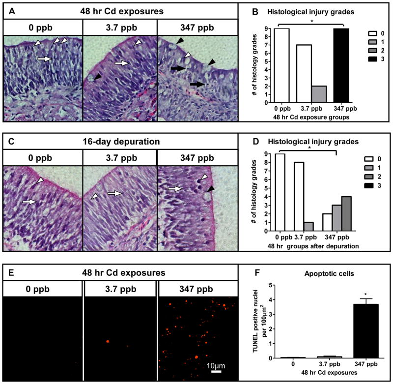
Fig. 2.
Histological effects of acute Cd exposure on the coho olfactory epithelium. (A and C) Light microphotograph (40× magnification) of cross-sectioned olfactory epithelium of coho exposed to 0, 3.7 and 347 ppb Cd for 48 hrs and following a 16 day depuration. SUS cells (white arrow heads), ORNs (white arrows), goblet cells (black arrowheads) and condensed nuclei (black arrows). (B and D) Histology injury grades of olfactory tissue sections following acute Cd exposures as represented in A and C. (E) Apoptosis in cross-sectioned olfactory epithelium of coho exposed to 0, 3.7 and 347 ppb Cd for 48 hrs (20× magnification). (F) Quantification of TUNEL positive nuclei per 100μm2 of olfactory epithelium. All data represent the mean ± SEM of n = 3 individuals. * indicates statistically significant differences in histological injury relative to controls (p <0.05).

