Fig. 3.

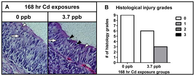
Histological effects of sub-chronic Cd exposure on the olfactory epithelium. (A) Light micrograph (40× magnification) of cross-sectioned olfactory epithelium of coho exposed to 0 and 3.7 ppb Cd for 168 hrs. SUS cells (white arrows), ORNs (white arrows), and goblet cells (black arrowheads). (B) Histology injury grades of olfactory tissue sections following sub-chronic Cd exposures represented in (A). All data represent the mean ± SEM of n = 3 individuals. * indicates statistically significant differences in histological injury relative to controls (p <0.05). (A).
