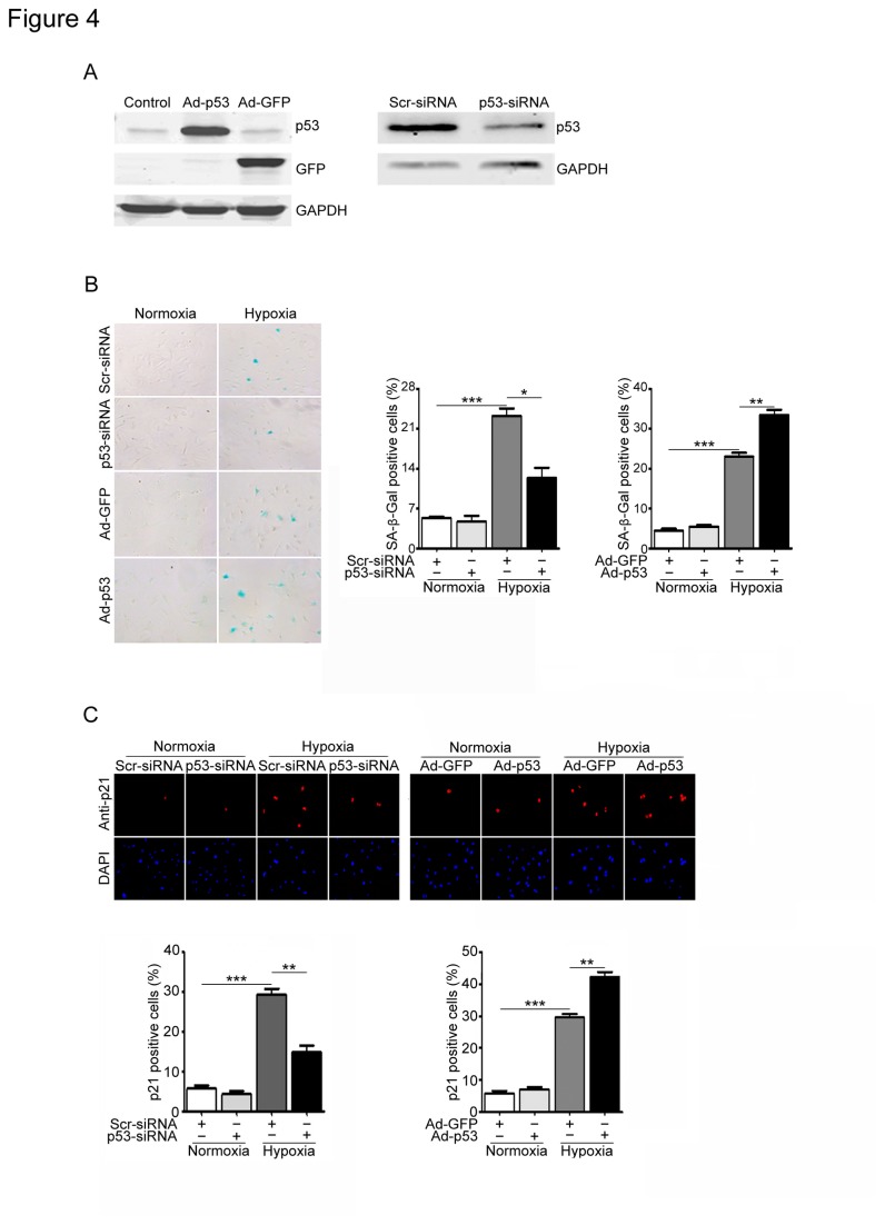
Figure 4. Effects of p53 on cell senescence and p21 expression in cardiac fibroblasts induced by hypoxia/reoxygenation.
(A) Cardiac fibroblasts were infected with scrambled siRNA (Scr-siRNA, 100 nmol/L), p53-siRNA (100 nmol/L), or adenovirus GFP control (Ad-GFP, MOI=50) or p53 (Ad-p53, MOI=50) for 24 h and then exposed to hypoxia/reoxygenation (H/R) for 3 days. The infection efficiency was detected by Western blot analysis using anti-p53 antibody. (B) Senescent cells were detected by SA-β-Gal staining (left). Bar graph shows the percentage of SA-β-Gal-positive cells (middle and right). (C) Senescent cells were subjected to immunostaining using anti-p21 antibody (top). DAPI was used for counterstaining (middle). Bar graph shows the percentage of p21-positive cells (bottom). Scale bars: 50 µm. Data expressed as mean±SEM (n=3). ***P<0.001 vs. normoxia; *P<0.05, **P<0.01 vs. Scr-siRNA+hypoxia or Ad-GFP+hypoxia.

