
Figure 6. Immune localization of LANA and histones in PEL cells.
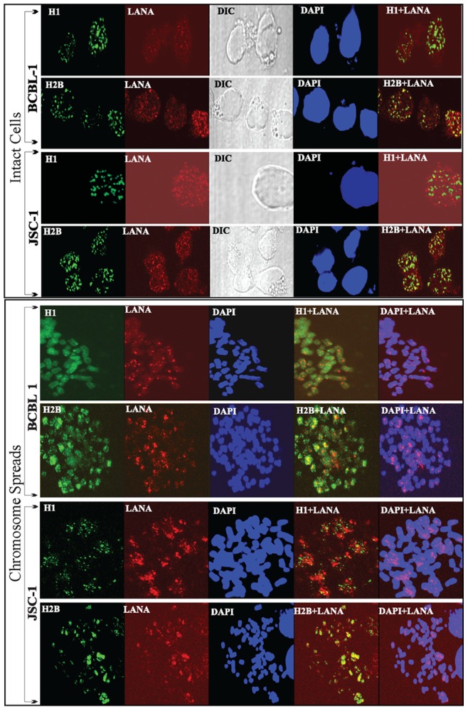
In the ‘Intact Cells’ panel, KSHV infected, BCBL1 and JSC1 cells were fixed on a glass cover slip before permeabilization with TritonX-100. Histone H1 and H2B were detected with mouse anti-H1 and Rabbit anti-H2B antibodies. LANA was detected with rat anti-LANA monoclonal antibody. Histone H1 and H2B were detected with AlexaFluor 488 (green) and LANA was detected with AlexaFluor 594 (red). Nuclei were stained with DAPI shown in blue. Image including DIC were captured using Olympus confocal microscope. In the ‘Chromosome Spreads’ panel, BCBL1 and JSC1 cells were treated with hypotonic solution to prepare chromosome spreads. These spreads on glass slides were fixed and permeabilized with TritonX-100. Histone H1 and H2B were stained with specific antibodies and were counter stained with AlexaFluor 488 (green) and LANA were detected with AlexaFluor 594 (red). Yellow signals in the merge panels show colocalization.