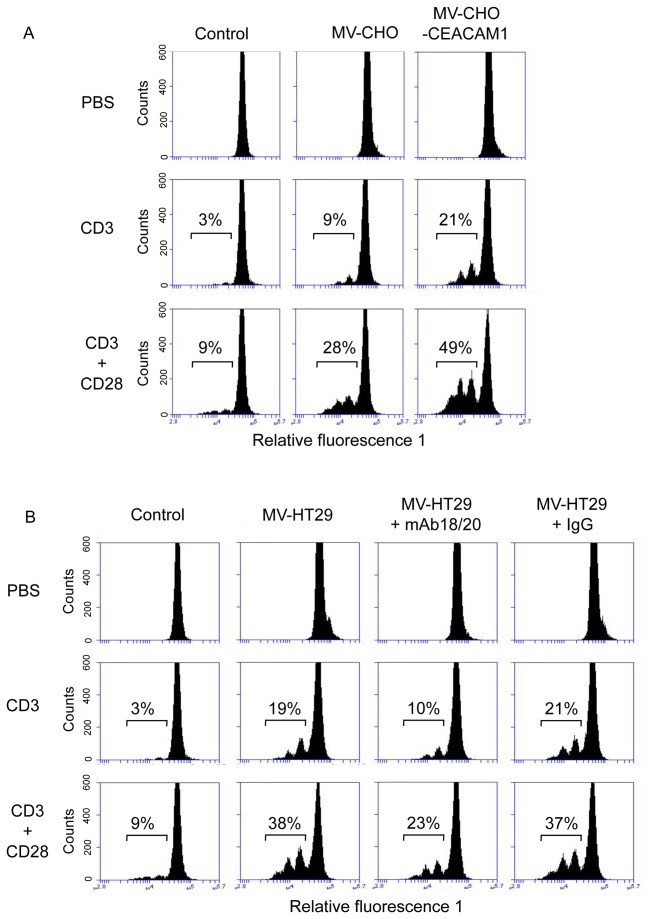
Figure 7. CEACAM1-positive MVs significantly increase the anti-CD3 and anti-CD3/CD28 mAb triggered T-cell proliferation.
Freshly isolated human PBMC were labeled with CFSE and cultured for 4 days in the presence of anti CD3 and anti CD3/CD28 with and without CHO- and CHO-CEACAM1 derived MVs (A). B) CFSE labeled PBMC were cultured for 4 days in the presence and absence of antiCD3 and antiCD3/CD28 with and without HT29-derived MVs. Untreated treated cells served as control. In indicated cases samples were co-cultured with antiCEACAM1 mAb18/20 (50 µg/ml) or isotype matched control IgG (50 µg/ml). Then PBMCs were analyzed utilizing the Accuri C6 flow cytometer system. The histograms depict PBMCs that have divided 1-3 times based on CFSE dilution peaks and reflex the cell proliferation rate given in %. The data shown are representative for three independent repeats of the experiment.

