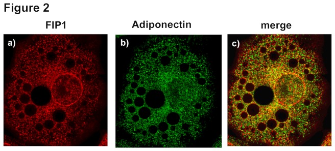
Figure 2. Intracellular distribution of FIP1, and adiponectin in adipocytes.

3T3L1 cells were cultured and differentiated to adipocytes as described in the methods section. Cells were fixed, permeabilized and immunostained with anti-FIP1 (Sigma) and anti-adiponectin (R and D) primary antibodies and Alexa-488 and Alexa-594 –conjugated secondary antibodies. Data shown is representative of cells obtained from three independent sets of cells. Determination of colocalization was carried out as described (n = 27).
