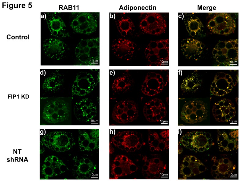
Figure 5. Knock Down of FIP1 does not impair traffic of adiponectin to the ERC compartment.
Control uninfected 3T3L1 cells or expressing non-targeting shRNAs or FIP1- shRNAs cultured and differentiated to adipocytes and used on day 10 after differentiation. Cells were fixed, permeabilized and stained with specific antibodies to detect endogenous rab11 and adiponectin proteins. Secondary antibodies conjugated to Alexa-488 or Alexa-594 respectively were used to visualize the proteins. Cells were visualized using a Leica DMI6000 microscope. Representative cells from 3 independent experiments are shown. Determination of colocalization was carried out as described (Control (n = 10), FIP1 shRNA (n = 9), NT shRNA (n = 4)). Panels c, f and i show the merged images.

