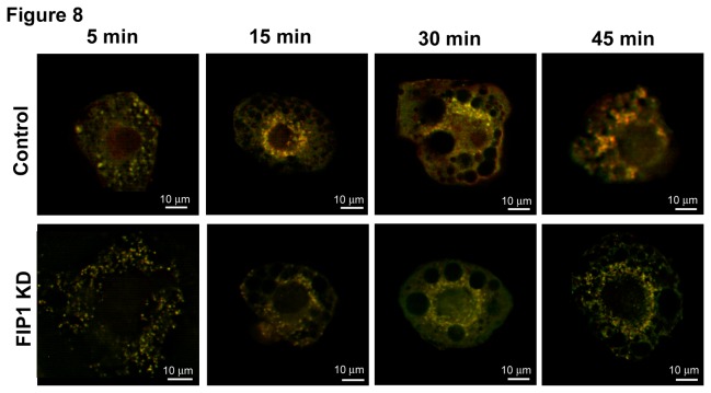
Figure 8. Adiponectin receptors are internalized and localize with transferrin receptor in intracellular membranes.
Control uninfected 3T3L1 cells or cells expressing shRNAs for FIP1 were cultured and differentiated to adipocytes and used on day 10 after differentiation. Cells were labelled with biotin-adiponectin and Alexa-594 conjugated transferrin at 4°C for 30 minutes and then incubated in Alexa488-streptavidin. Internalization of adiponectin and transferrin bound receptors was allowed at 37°C for the times as indicated. Cells were then transferred to ice, and fixed in PFA as detailed in the methods section. Cells were imaged in a Leica DMI6000 microscope. Data shown are the merged images. Representative cells are shown selected from three independent experiments.

