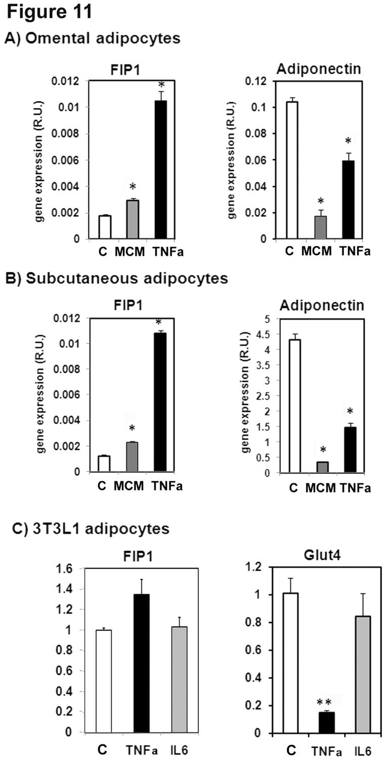
Figure 11. Pro-inflammatory cytokines increase the expression of FIP1 in adipocytes.

Human preadipocyte cells from omental (A) or subcutaneous (B) adipose tissue were obtained from Zen-Bio and differentiated as per their instructions for 14 days. A subset of cells were left untreated (control, C) or were incubated in media supplemented with macrophage conditioned media (MCM, 5% v/v) or with 100 ng/ml of Tumor necrosis factor alpha (TNFα), Total RNA was isolated and FIP1 and adiponectin mRNA was quantitated by real time PCR as indicated in the methods section. Relative quantification was carried out using the ∆∆Ct method using PPIA gene expression as an internal control. Statistical analysis: One way ANOVA. * indicates statistical significance p<0.05. Data from one experiment with triplicate cell dishes. C) Fully differentiated 3T3L1 adipocytes were incubated in the absence (control, C) or presence of TNFα (TNFα) or Interleukin 6 (IL6) at 50 ng/ml for 24 hours, Total RNA was isolated and the amount of mRNA for FIP1, or Glut4 was determined by real time PCR. Quantification was carried out using the ∆∆Ct method using cyclophyllin A as internal control. The graph shows data from a representative experiment of two independent experiments each carried out with triplicate cell dishes for each group. Statistical analysis: One way ANOVA, ** indicates p<0.01.
