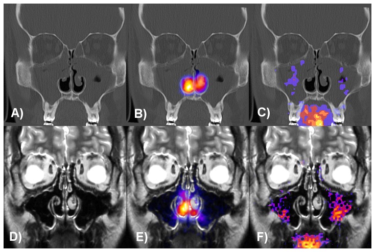
Figure 4. Coronal CT and MRI slices of a CRS patient before and 166 days after sinus surgery.
Superposition of the anterior gamma camera images with the CT slice before FESS (A–C) without (B) and with (C) central nasal lead shield mask and with the MRI slice after FESS (D–F) without (E) and with (F) lead shield. Lund-Mackay score was 15 and maxillary sinus deposition was 1.3% (C) before and 7.9% (F) after surgery. The patient had septoplasty and conchotomy on both sides eight years before participation in the study.

