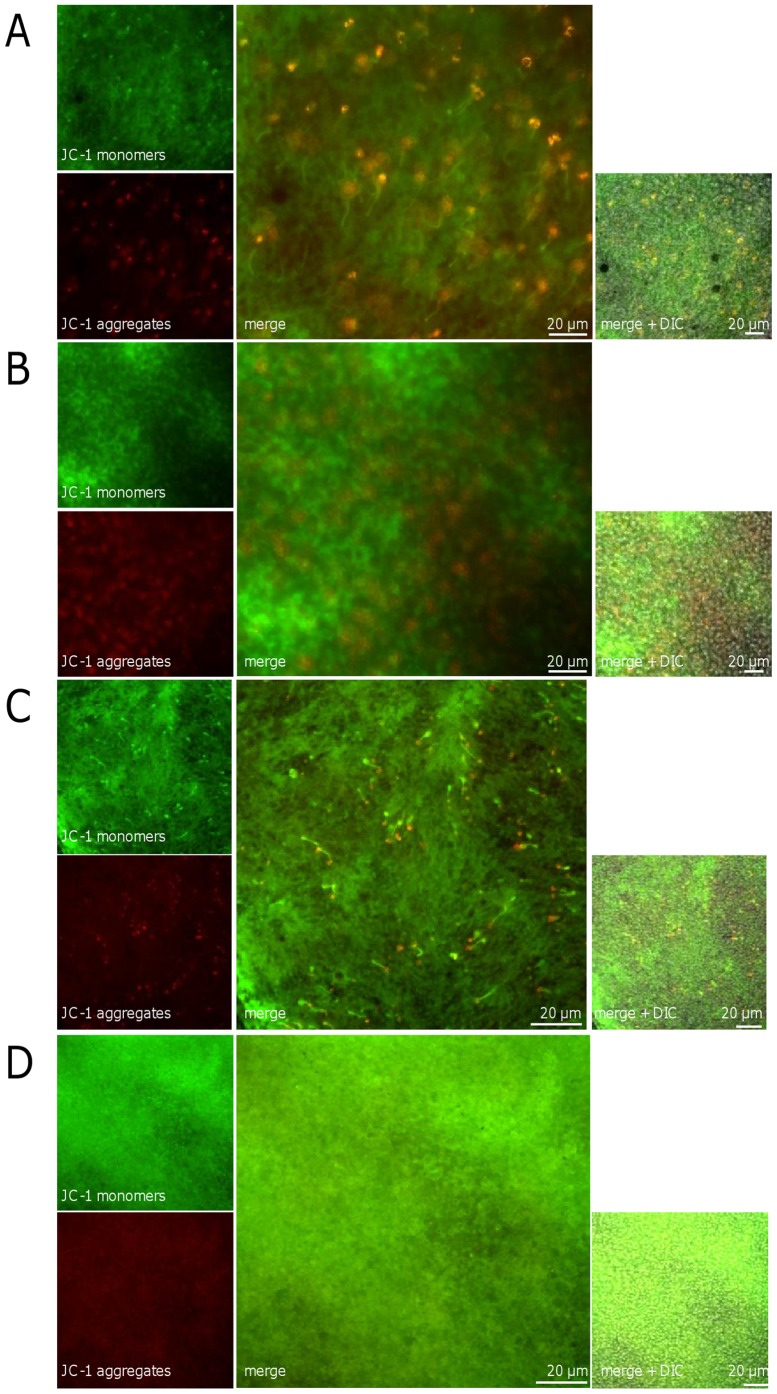
Figure 12. Shift of JC-1 staining after blue light irradiation in OS.
Images of the outer segment layer of whole mounts stained with 10 µg/ml JC-1 after cultivation. The eyes were irradiated with blue light for 6 h (B) and 12 h (D), the time-matched controls cultivated for 6 h (A) and 12 h (C), respectively. An increase of green JC-1 monomers (sign for MMP collapse) was detected in both irradiated retinas to a greater extent than in the controls. Some fluorescent red J-aggregates (seen in healthy cells) were still visible after 12 h irradiation (D). In the controls the aggregates were observed as spots additionally to the staining of the outer segments in the whole mount (B, D). Images are representative of three experiments.

