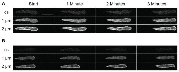
Figure 4. Crawling cells can advance with only minor snaking.
Two cAR1-GFP expressing cells (A, B) crawling toward a cAMP source. Fluorescence images taken as in Figure 1 and presented at 1 minute intervals. Three images for each time point are shown: these capture the base of the cell on the coverslip (cs), and 1 µm or 2 µm above that. Images were taken each 5 seconds; the complete data set can be seen as Video S1 and Video S2 in Supporting Information. The cells move with little lateral motion except at the front of each cell.

