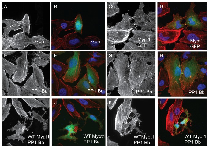
Figure 5. PP1Ba and PP1Bb assemble myosin phosphatase complexes that regulate the actin cytoskeleton.
HeLa cells were transfected with either GFP alone (A, B), zebrafish Mypt1 (1-300) and GFP (C, D), zebrafish GFP-PP1Ba (E, F), zebrafish GFP-PP1Bb (G,H), Wild-type Mypt1 and PP1Ba (I, J), wild-type Mypt1 and PP1Bb (K, L). All cells were fixed and stained with DAPI and Alexa 568-phalloidin and imaged with confocal microscopy. Black and white images show phalloidin staining, while color images are a merge of DAPI (blue), GFP (green) and phalloidin (red).

