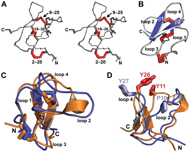
Figure 7. Structure of OAIP-1.
(A) Stereoview of the ensemble of 20 OAIP-1 structures. The three disulfide bonds and the N- and C-termini are labeled. (B) Schematic (Richardson) representation of OAIP-1. β-strands are colored blue and disulfide bonds are shown as red tubes. The four intercystine loops (loops 1–4) are labeled. (C) Overlay of OAIP-1 (blue) and the orthologous toxin U1-TRTX-Pc1a (orange). The intercystine loops and termini are labeled. (D) Overlay of OAIP-1 (blue) and the orthologous toxin U1-TRTX-Pc1a (orange). Residues Y11 and Y26 in U1-TRTX-Pc1a (red tubes) interact in such a way that loops 2 and 4 are brought into close proximity. The equivalent residues in OAIP-1, P10 and Y27 (light blue tubes), do not interact and consequently loops 2 and 4 are further apart.

