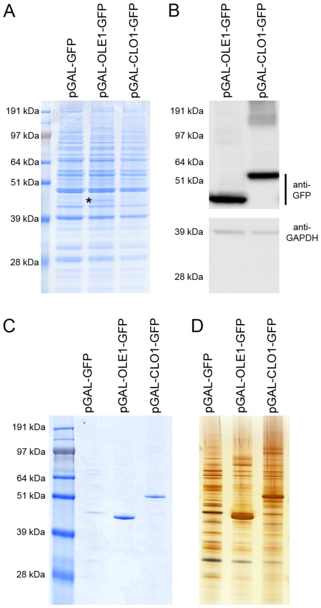
Figure 1. Expression of AtOle1-GFP and AtClo1-GFP in yeast.

Total protein extracts from GFP, AtOle1-GFP and AtClo1-GFP expressing cells were analyzed using SDS-PAGE (A) and immunoblots (B). The presence of AtOle1-GFP (*) was visible on the Coomassie blue stained gel and the fusion proteins were also detected using an anti-GFP antibody. The anti-GAPDH antibody was used as a loading control. Association of the plant proteins with yeast lipid droplets was confirmed by SDS-PAGE analysis of the protein profile of purified lipid droplets. The proteins were revealed by Coomassie blue staining (C) or silver staining (D).
