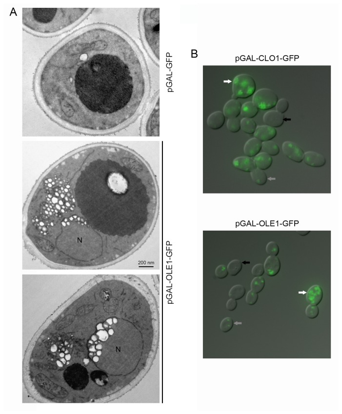
Figure 3. Observation of yeast cells expressing plant oleosin and caleosin.
BY cells transformed with pGAL-GFP, pGAL-OLE1-GFP or pGAL-CLO1-GFP were cultured for 18 h in a galactose-containing medium. Cells were processed for electron microscopy (A). Lipid droplets appear as white round structures surrounded by a black membrane. N, nucleus. Cells were also observed for detection of GFP signal using fluorescence microscopy (B). White, grey and black arrows indicate cells with high, medium or no plant protein expression, respectively.

