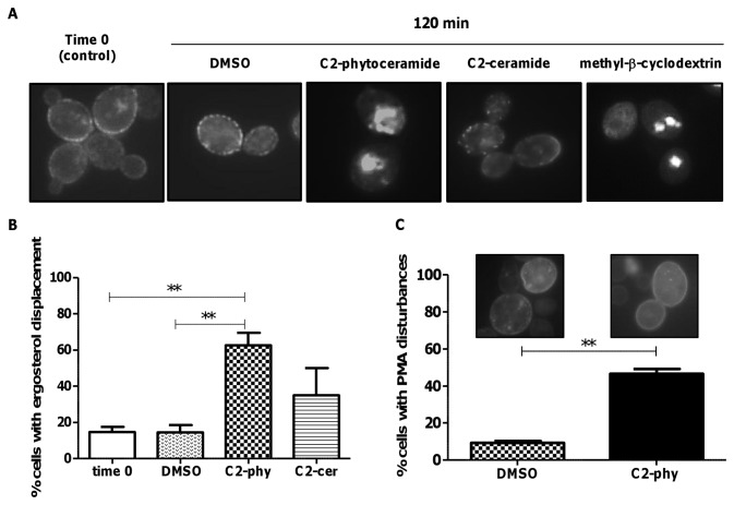
Figure 5. Distribution of sterol-rich domains in S. cerevisiae cells exposed to C2-phytoceramide.
(A) Fluorescence microscopy pictures of W303-1A cells exposed to 30 µM C2-phytoceramide, 40 µM C2-ceramide, 5 mg/ml methyl-®-cyclodextrin or 0.1% DMSO for 120 min and stained with filipin (5 mg/ml). (B) Percentage of yeast cells with ergosterol displacement. Cells were treated as described in (A) and the number of cells with ergosterol displacement was determined by counting at least 120 cells per sample, in three independent experiments. P<0.01 respectively, One-Way ANOVA. (C) Percentage of yeast cells with perturbed Pma1p-GFP distribution. W303-1A cells were transformed with a single copy vector derived from pRS316 expressing Pma1p-GFP (3). At least 300 cells per sample were counted. P<0.01.

