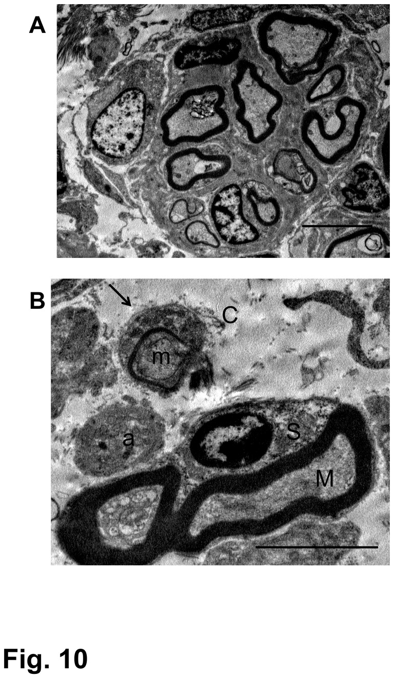
Figure 10. Electron microscopy of regenerating nerve fibers.
These micrographs were obtained from the spinal cord epicenter of the rat that had received BMSC transplantations starting at 4 weeks after contusion injury and was fixed 4 weeks following the initial cell injection (4-w PI). This rat showed a BBB improvement from 4 points before cell injection to 10 at fixation.
(A) Myelinated nerve fibers are grouped in a small fascicle, which is surrounded by a clear, cell-free space. Scale: 5 µm.
(B) Part of the nerve fascicle is enlarged. There are thick-myelinated (M) and thin-myelinated (m) axons. The former is surrounded by a Schwann cell (S), and the latter is associated with a thin Schwann cell cytoplasm, the basal lamina of which is clearly seen (arrow). An unmyelinated fiber (a) is also seen near these myelinated axons. These nerve fibers are located in the space, through which collagen fibrils (C) are distributed, partly in association with Schwann cells. Scale: 5 µm.

