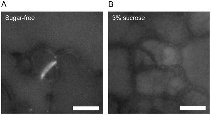
Figure 4. Aniline blue staining of cotyledon epidermal cells.
Four day-old cotyledons immersed in sugar-free (A) or 3% sucrose (B) solutions were stained with 0.02% aniline blue for 1 week. Representative images from 24 (sugar-free) and 38 (3% sucrose) independent seedlings were shown. Note that aniline blue fluorescence was clearly detected in new cell walls forming in meristemoids immersed in sucrose-free solutions but not in 3% sucrose solutions. Scale bars = 10 μm.

