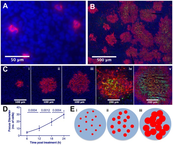
Figure 1. Extravasation and spatially constrained spread of systemic oncolytic therapy.
Immunofluorescence analysis and quantification of 5TGM1 tumors harvested following IV VSV administration, sectioned and stained to detect VSV (red), dying cells (TUNEL, green) and tumor cell nuclei (Hoescht, blue). (A) Seeds of infection established following virus extravasation 24 hr post-VSV(ΔG). (B) Expansion and conflation of intratumoral foci and destruction of tumor cells 48 hr post-VSV. (C) Radial expansion of infection and subsequent cell death of intratumoral focus in tumor harvested at 6, 12, 18, 24, and 48 hr post-VSV(i-v). (D) Quantification of mean infectious focus diameter (n = 7–9/interval) in tumors harvested at 6 hr intervals post-VSV. (E) Schematic representation of proposed model of systemic oncolytic therapy showing (i) extravasation and infection of tumor cells seeding randomly distributed infectious centers, (ii) spatially constrained expansion, and (iii) conflation of foci resulting in viral destruction of tumor cells, though voids of uninfected, surviving cells remain.

