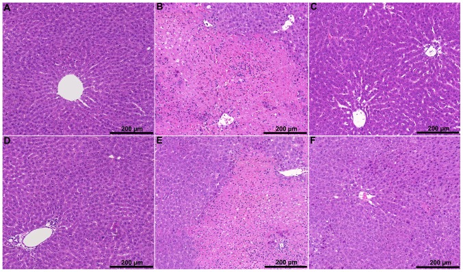
Figure 4. Representative H&E-stained liver sections.
In the control groups (A: “early” control; D: “late” control) mild tissue injury and sinusoidal dilatation were observed. In the IR groups (B: “early” IR; E: “late” IR) confluent necrotic areas were detected accompanied by significant leukocyte infiltration and tissue hemorrhage. The levosimendan pretreated groups (C: “early” levosimendan pretreatment; F: “late” levosimendan pretreatment) were characterized by focal necrosis associated with milder tissue hemorrhage and less severe leukocyte infiltration.

