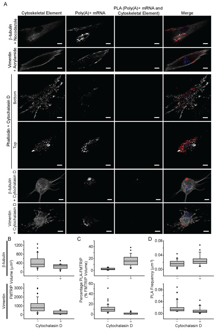
Figure 2. Interactions between poly(A)+ mRNA and cytoskeletal elements in HDF post-depolymerization of microtubules using nocodazole, intermediate filaments using acrylamide, and actin using cytochalasin D.
(A) β-tubulin, vimentin, and phalloidin immunofluorescence (IF), poly(A)+ mRNA, and PLA between poly(A)+ mRNA and the cytoskeletal elements in HDF were imaged with a laser-scanning confocal microscope. Merged images of the cytoskeleton (white), poly(A)+ mRNA (red), PLA (green) and nuclei (blue) are shown. Single image plane is represented. Scale bar, 10 µm. (B) The mean FMTRIP volume decreased after 90 min exposure to cytochalasin D (Mann-Whitney rank sum test, β-tubulin, p<0.01; vimentin, p<0.01) in experiments quantifying interactions with β-tubulin (n=21, mean=254µm3, s.d.=110µm3) and vimentin (n=16, mean=253µm3, s.d.=188µm3). (C) The mean percentage of FMTRIP (PLA-FMTRIP) bound to β-tubulin increased after depolymerization (Mann-Whitney rank sum test, p<0.001; n=21, mean=16.3%, s.d.=9.5%) but decreased for vimentin (Mann-Whitney rank sum test, p<0.001; n=16, mean=1.5%, s.d.=1.9%). (D) The mean PLA frequency detecting interactions with β-tubulin also increased (Mann-Whitney rank sum test, p=0.015; n=21, mean=0.03µm-3, s.d.=0.02µm-3) and decreased for vimentin (Mann-Whitney rank sum test, p<0.001; n=16, mean=0.01µm-3, s.d.=0.01µm-3). Error bars, s.d.

