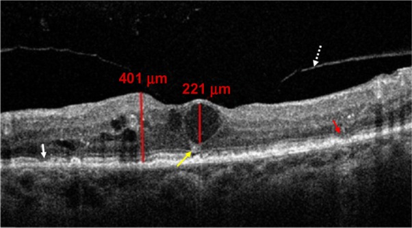
Figure 2.

OCT scan showing CME II D+ with disruption of ELM (red arrow) and IS/OS junction line (white arrow). Hyper-reflective foci (yellow arrow) and partial PVD without traction (white dotted arrow) are also shown.
Abbreviations: CME, cystoid macular edema; OCT, optical coherence tomography; ELM, external limiting membrane; IS/OS, inner segment/outer segment; PVD, posterior vitreous detachment.
