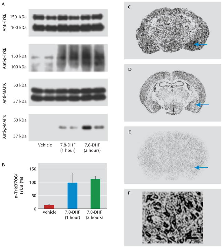
FIGURE 1. Activation of p-TrkB and MAPK in Mouse Amygdala by Systemic 7,8-Dihydroxyflavone (7,8-DHF)a.
a MAPK=mitogen-activated protein kinase. Panel A shows immunoblots of mouse amygdala punches examining total TrkB protein (top), activated p-TrkB (second row), total MAPK (third row), and activated p-MAPK (bottom row). Each condition is represented in duplicate, with amygdala punches from mice injected intraperitoneally with vehicle (control) or 7,8-DHF (5 mg/kg) 1 hour or 2 hours prior to sacrifice. Full-length TrkB is detected at both ~95 kDa (nonglycosylated) and ~140–145 kDa (glycosylated) forms. Although total levels of TrkB and MAPK do not change, systemic 7,8-DHF led to robust activation/phosphorylation of amygdala TrkB (p-TrkB Y706) and MAPK (p-MAPK). Panel B shows the quantification (mean values with standard deviations) of the p-TrkB706:TrkB ratio represented in the top two immunoblots of panel A, demonstrating increased p-TrkB706 with 7,8-DHF treatment. In panel C, receptor autoradiography demonstrates that 3H-7,8-DHF binds to brain regions known to express TrkB protein, including the amygdala (arrow). Panel D shows in situ hybridization of TrkB mRNA expression from the Allen Brain Atlas (www.allenbrainatlas.com). In panel E, 3H-7,8-DHF shows no significant binding when tissue is pretreated with excess cold 7,8-DHF, suggesting relative specificity. Panel F shows dense expression of TrkB protein on pyramidal neurons in basolateral rat amygdala identified with immunocytochemistry. Scale bar represents ~50 μm.

